Home

Rosée demander préféré tampon x ray Pickering fuite pour

Tampons de gaze Setpack® RK de Lohmann & Rauscher - farla medical - Promo 2%

The truth about correctional facility backscatter X-ray body scanners

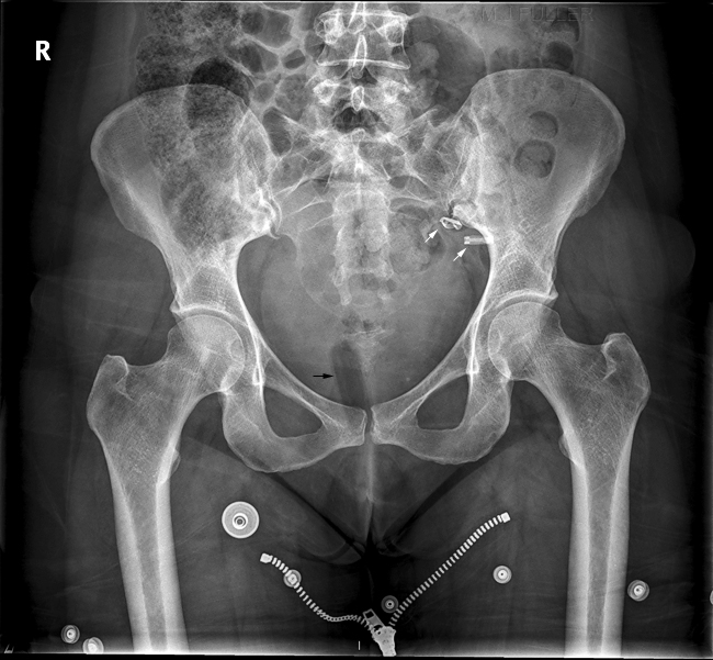
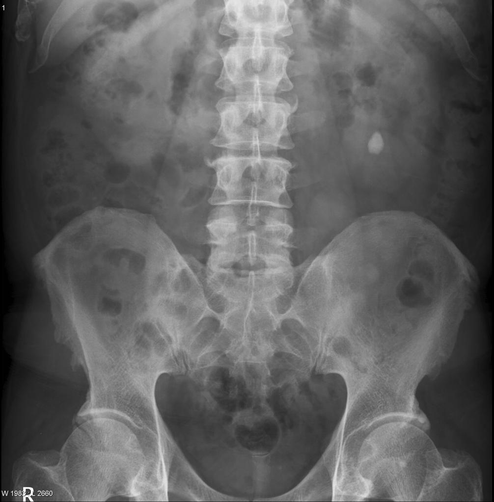
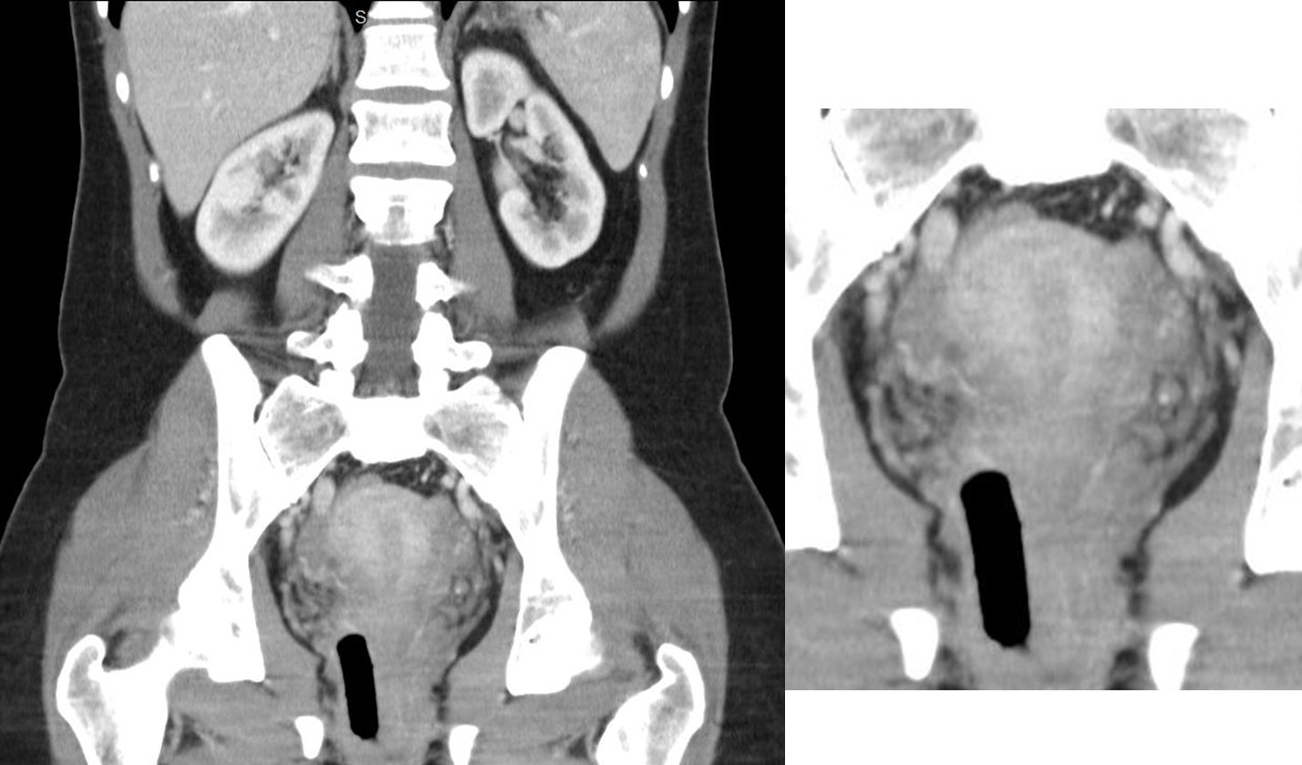
Tampon visible on hip radiograph | Radiology Case | Radiopaedia.org

Tampon Appearance on Bone Scan Imaging: Case Report - ScienceDirect

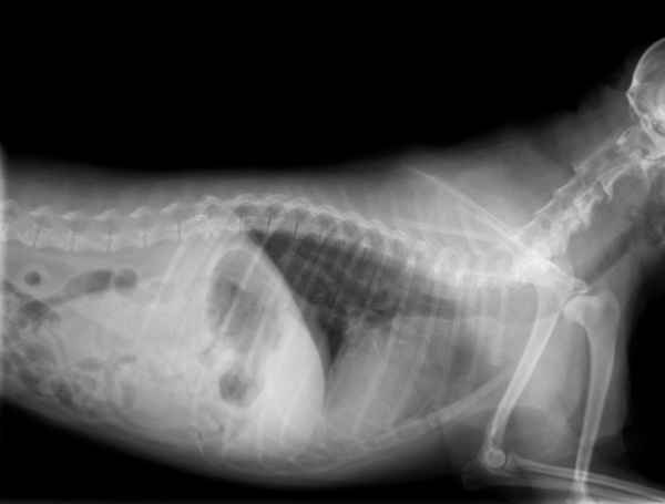
Your Dog Ate a Tampon? Act Fast: Our Vet Explains

Vaginal Foreign Body Evaluation and Treatment Article

How to insert a naso‐enteric tube using an ultrathin scope - Shukla - 2021  - ANZ Journal of Surgery - Wiley Online Library
How to insert a naso‐enteric tube using an ultrathin scope - Shukla - 2021 - ANZ Journal of Surgery - Wiley Online Library

Abdominal x rays made easy: iatrogenic, accidental, and incidental objects  | The BMJ
Abdominal x rays made easy: iatrogenic, accidental, and incidental objects | The BMJ

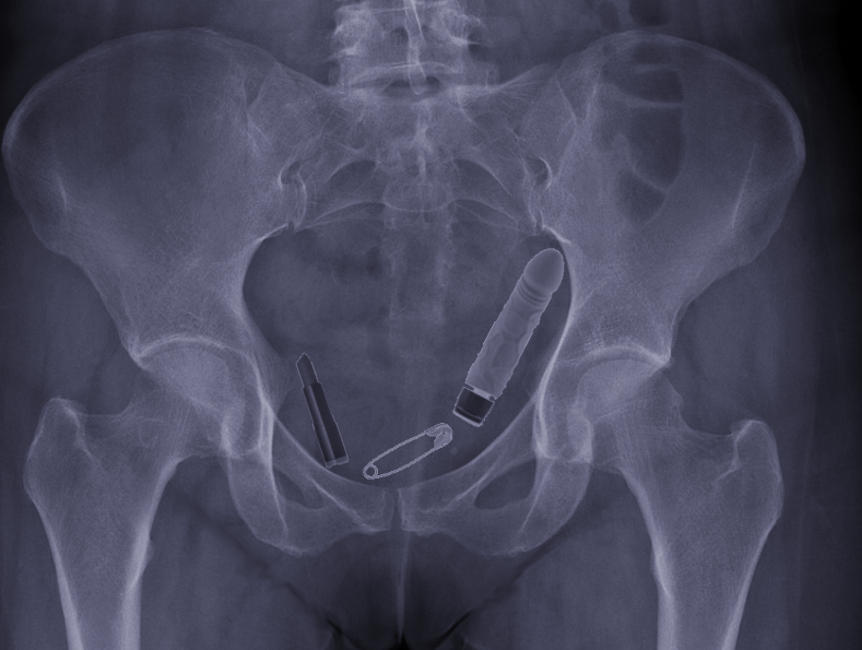
Foreign bodies of habitual use found in the vaginal cavity in the x-ray, CT  scan and MRI | Semantic Scholar
Foreign bodies of habitual use found in the vaginal cavity in the x-ray, CT scan and MRI | Semantic Scholar

Abdominal x rays made easy: iatrogenic, accidental, and incidental objects  | The BMJ
Abdominal x rays made easy: iatrogenic, accidental, and incidental objects | The BMJ

Radiology case: Tampon on plain radiograph

2014 X-Ray Contest Winners - Veterinary Practice News

Tampon in-situ | Radiology Case | Radiopaedia.org

Pelvis Anatomy, Artifacts and Variants - wikiRadiography

Plain X-ray with a U shape device in the pelvis minor and without any... |  Download Scientific Diagram
Plain X-ray with a U shape device in the pelvis minor and without any... | Download Scientific Diagram

Sterile X-Ray Detectable Tampon 7.5 x 2.8cm | Vernacare

a) Pelvic radiograph shows a typical IUD (arrowhead). A tampon (arrow)... |  Download Scientific Diagram
a) Pelvic radiograph shows a typical IUD (arrowhead). A tampon (arrow)... | Download Scientific Diagram

Tampon de gaze - X-RAY and LOOP - Albino Dias de Andrade S.A. - en coton

a) Pelvic radiograph shows a typical IUD (arrowhead). A tampon (arrow)... |  Download Scientific Diagram
a) Pelvic radiograph shows a typical IUD (arrowhead). A tampon (arrow)... | Download Scientific Diagram

The Abdominal XRay: A relic or a reliable tool? — Taming the SRU

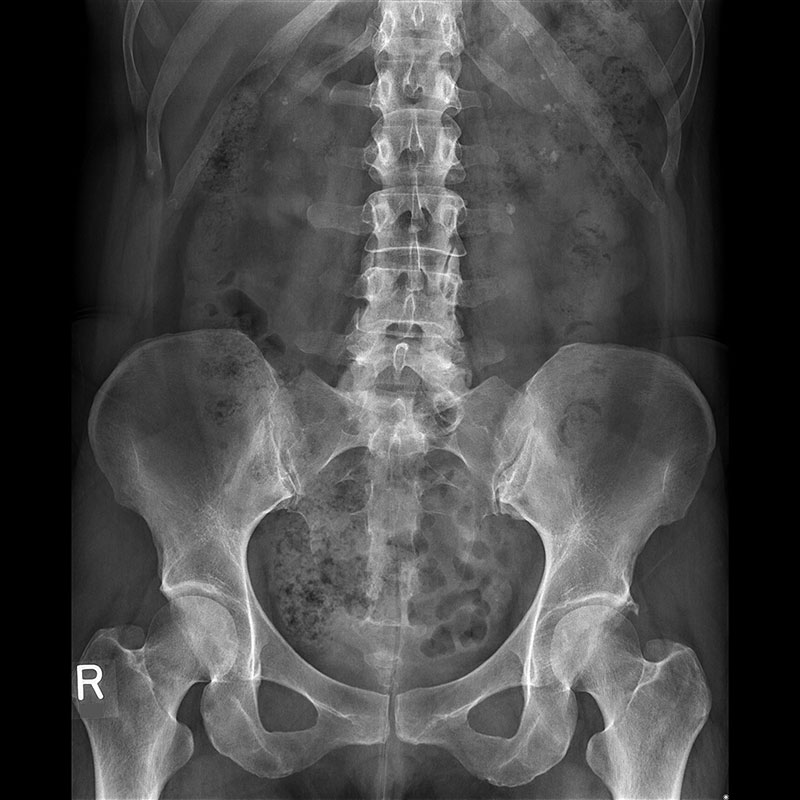
File:Tampon im Roentgenbild des Beckens 45W - CR ap - 001 - Annotation.jpg  - Wikimedia Commons
File:Tampon im Roentgenbild des Beckens 45W - CR ap - 001 - Annotation.jpg - Wikimedia Commons

Premier Detectors Premier Detectors Sterile X-Ray Detectable Tampons No.1  (2.5 x 7.5 cm)
Premier Detectors Premier Detectors Sterile X-Ray Detectable Tampons No.1 (2.5 x 7.5 cm)

Radiopaedia.org - 25yo female. What is the foreign body seen in the pelvis?  ANSWER: https://goo.gl/3AxDLx Case courtesy of Dr Vikas Shah. | Facebook
Radiopaedia.org - 25yo female. What is the foreign body seen in the pelvis? ANSWER: https://goo.gl/3AxDLx Case courtesy of Dr Vikas Shah. | Facebook

EPOS™
EPOS™

Ultimax-i X-ray Clinical Gallery | Canon Medical Systems USA

Filthy Foreign Bodies - Veterinary Emergency & Surgery Hospital

Tampon on abdominal x-ray | Radiology Case | Radiopaedia.org